EP – Evoked Potentials
What are Evoked Potentials?
During these tests, a specific stimulus is repeatedly given. On the head or spine, the electrical response of the brain or spinal cord to this stimulus is recorded and isolated from other electrical activities of the brain. The electrode is placed precisely over the brain region where these stimuli can be perceived. With this simple method, interruptions or damage to the nerve pathways in our body can be measured. Evoked potentials are signals with low amplitude compared to spontaneous potentials (e.g., EEG). To record them, the stimuli must be repeatedly presented.
Visual Evoked Potentials (VEP) test the visual pathways
These allow for the measurement of the conduction of nerve impulses through the visual pathway. The visual pathway is stimulated using a checkerboard pattern that changes its black-and-white profile at fast, regular intervals. The patient observes the pattern through a screen with one eye. The electric potentials generated in the visual cortex are recorded by the doctor using electrodes attached to the patient’s head.
This measurement method is relevant, among other things, in diagnosing inflammatory or circulatory changes in the optic nerve and visual pathway (e.g., optic neuritis in multiple sclerosis).
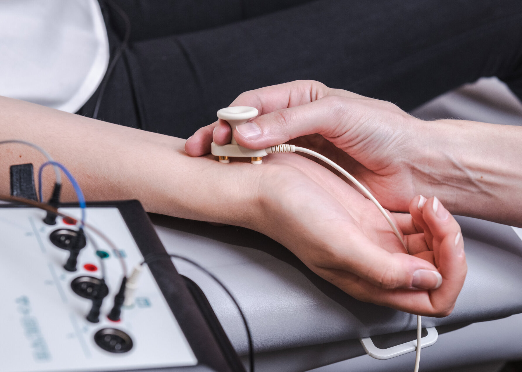
Somatosensory Evoked Potentials (SSEP) test the pathways that convey touch sensations
The SSEP enables an evaluation of the central and peripheral sensory pathways. With the SSEP, the doctor can test the transmission of sensory perceptions from the arms and legs through the spinal cord to the brain. This is done by repeatedly triggering short electrical stimuli at the ankles and wrists. The signals generated are measured over the head or spinal cord.
This method is used, for example, to more accurately assess sensory disturbances (e.g., in multiple sclerosis).
Motor Evoked Potentials (MEP) test the pathways that convey movements
The MEP examines the transmission of nerve impulses responsible for the transmission of muscle movements from the brain through the spinal cord to the extremities (arms and legs).
During the examination, a strong magnetic field is applied over the patient’s head, neck, or lumbar spine, which stimulates corresponding areas of the cerebral cortex or spinal cord. This results in brief, involuntary muscle contractions in the arms or legs, depending on the stimulated brain area. Some patients may find them unpleasant, but they are not dangerous (some rare contraindications are known to the doctor and should be observed).