EEG – Electroencephalography
What is an Electroencephalogram (EEG)?
The EEG is a method for measuring the electrical activity of the brain. The natural voltage fluctuations of the cerebral cortex are recorded through this method.
How does the EEG work?
Metal plates (electrodes) are placed on the scalp (usually 21). The electrical voltage differences between each pair of electrodes are measured in various combinations, amplified with a special device, and finally recorded as brain wave patterns. These electrical voltages are generated by the nerve cells themselves and are a sign of their activity and functionality.
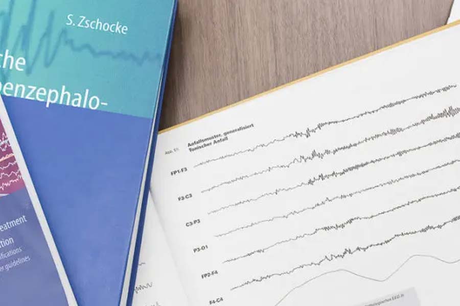
The doctor evaluates the brain wave patterns based on their frequency, wave height (amplitude), steepness, and location on the surface of the brain. Symmetry between the two brain hemispheres is also important.
What is the EEG used for?
An EEG is used to detect pathological changes in the electrical activity of the brain. These include:
- Epilepsy-specific potentials: These are typical EEG changes that can occur with a tendency to have epileptic seizures (convulsions of the brain). The EEG can help diagnose epilepsy. However, the EEG often does not show any abnormalities between seizures. Therefore, it may need to be performed multiple times and under conditions that increase seizure susceptibility (e.g., sleep deprivation the night before the examination).
- General changes in EEG findings: These affect the entire cerebral cortex and can occur, for example, with inflammatory (encephalitis) or metabolic brain diseases or poisonings. They can provide a clue about the severity of the disease.
- Focal findings: These are regional changes in brain wave patterns that occur mainly in local brain diseases such as stroke, tumor, or local inflammatory processes.
Today, the EEG is most commonly used for diagnostic purposes when there is suspicion of an epileptic event.
How is the EEG performed?
Usually, 21 electrodes are evenly placed on the patient’s scalp to record the brain waves. To prevent them from moving, the electrodes are attached to a cap that spans the forehead and hairy parts of the head like a hairnet.
A routine EEG takes about 30 minutes. Sometimes this resting measurement is supplemented with a flicker light examination with closed eyes. The patient must look at a rapid light-dark sequence (stroboscope). This can reveal previously hidden epileptic tendencies. In special cases, an EEG measurement over 24 hours may be necessary. The EEG examination is harmless, painless, and can be repeated as often as desired.